
Spinal Decompression Surgery
 Evaluation and surgical treatment for spinal stenosis, herniated discs, nerve compression, sciatica, arm numbness, walking limitations, and spinal cord compression symptoms.
Spinal decompression surgery is performed to relieve pressure on spinal nerves or the spinal cord when compression causes significant pain, numbness, weakness, or loss of function.
For some patients, symptoms begin gradually with neck pain, lower back pain, or radiating arm and leg symptoms. Others develop progressive numbness, weakness, walking intolerance, balance problems, or difficulty performing normal daily activities.
When nerve compression continues despite conservative treatment, surgical evaluation may help determine whether decompression surgery is appropriate.
Dr. Garges provides spinal decompression surgery evaluation in Webster, TX for patients throughout Clear Lake, League City, Nassau Bay, Friendswood, Pearland, Pasadena, and the greater Houston area.
As a board-certified orthopedic spine surgeon with fellowship training at the San Francisco Spine Institute and prior experience as chief of spinal surgery at UTMB, Dr. Garges evaluates cervical, thoracic, and lumbar spine conditions requiring advanced surgical decision-making.
What Spinal Decompression Surgery Is Designed to Treat
Spinal decompression surgery is intended to relieve pressure on compressed nerves or the spinal cord caused by structural narrowing within the spine.
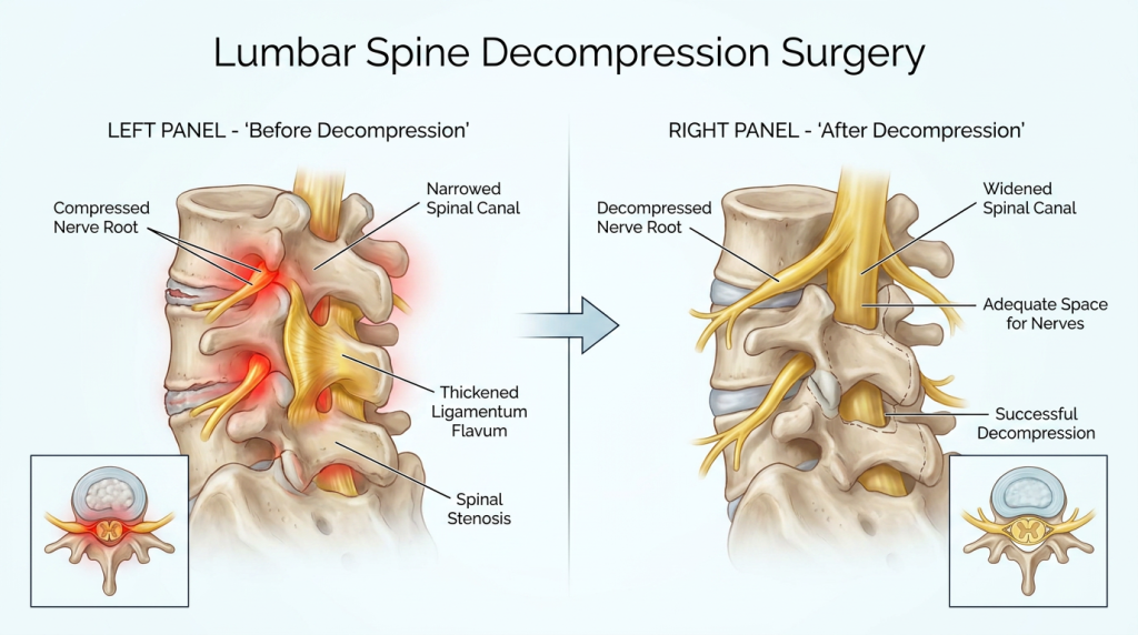
Nerve compression may develop from:
- Spinal stenosis
- Herniated discs
- Bone spurs
- Degenerative disc disease
- Thickened spinal ligaments
- Foraminal narrowing
- Degenerative instability or spondylolisthesis
Compression affecting the cervical spine may cause neck pain, arm symptoms, hand weakness, coordination difficulty, or spinal cord dysfunction.
Compression affecting the lumbar spine commonly causes lower back pain, sciatica, leg numbness, weakness, standing intolerance, or reduced walking capacity.
The goal of decompression surgery is to create additional space around compressed nerves or the spinal cord contributing to pain, neurologic symptoms, and functional limitations.
Not all spinal abnormalities seen on MRI require surgery. Surgical decisions are based on the relationship between symptoms, neurologic findings, physical examination, imaging studies, and response to prior conservative treatment.
Symptoms That May Indicate Significant Nerve Compression
Patients often seek evaluation when symptoms begin interfering with normal daily function, mobility, work activities, or quality of life.
Common symptoms associated with spinal nerve compression include:
- Persistent neck pain or lower back pain
- Sciatica or pain radiating into the leg
- Arm pain, numbness, or tingling
- Weakness in the hands, arms, legs, or feet
- Burning pain between the shoulder blades
- Difficulty standing or walking for extended periods
- Loss of balance or coordination
- Difficulty holding or gripping objects
- Symptoms disrupting sleep
- Functional decline affecting work or daily activities
Some patients experience progressive walking limitations caused by lumbar spinal stenosis. Others develop worsening hand coordination, dropping objects, or balance difficulty related to cervical spinal cord compression.
Symptoms may worsen while standing, walking, driving, bending, lifting, or maintaining certain neck positions. In some cases, patients notice gradual neurologic decline over time.
Because symptoms may overlap with shoulder, hip, peripheral nerve, or other musculoskeletal conditions, accurate diagnosis is important before surgical decisions are made.
Conditions Commonly Evaluated for Spinal Decompression Surgery
Several spine conditions may lead to compression of spinal nerves or the spinal cord. Surgical decompression may become a treatment consideration when conservative care no longer provides sufficient relief or when neurologic symptoms progress.
Spinal Stenosis
Spinal stenosis refers to narrowing of the spinal canal or nerve passageways. Degenerative changes associated with aging commonly contribute to stenosis.
Bone overgrowth, thickened ligaments, bulging discs, and degenerative arthritis may narrow the spinal canal and compress neural structures.
Lumbar spinal stenosis commonly causes:
- Leg pain while walking
- Standing intolerance
- Sciatica
- Leg weakness or numbness
Cervical spinal stenosis may contribute to:
- Arm numbness or weakness
- Hand coordination problems
- Balance changes
- Spinal cord compression symptoms
Herniated Discs
A herniated disc occurs when disc material extends beyond its normal boundary and compresses nearby nerves.
Cervical disc herniation may cause neck pain with radiating arm symptoms. Lumbar disc herniation commonly causes sciatica, leg pain, numbness, or weakness.
Some herniated discs improve with conservative care alone. Others continue causing persistent neurologic symptoms despite therapy, medications, injections, or activity modification.
Learn more about Herniated Disc Treatment.
Foraminal Stenosis and Nerve Root Compression
Foraminal stenosis refers to narrowing of the openings where spinal nerves exit the spine. This narrowing may compress individual nerve roots and produce radiating pain, numbness, or weakness.
Degenerative changes, bone spurs, disc collapse, and arthritis commonly contribute to foraminal narrowing.
Degenerative Instability and Spondylolisthesis
Some patients develop spinal instability in addition to nerve compression. Degenerative spondylolisthesis occurs when one vertebra shifts forward relative to another.
This instability may contribute to spinal canal narrowing and worsening neurologic symptoms during standing or walking.
How Dr. Garges Evaluates Surgical Candidacy
Not every patient with spinal stenosis or disc herniation requires surgery. Determining whether decompression surgery may help requires careful correlation of symptoms, imaging findings, neurologic examination, and prior treatment response.
Dr. Garges performs a comprehensive surgical evaluation that may include:
| Evaluation Component | Purpose |
|---|---|
| Symptom Review | Determines pain patterns, walking limitations, numbness, weakness, and progression of symptoms |
| Neurologic Examination | Evaluates strength, reflexes, sensation, coordination, and spinal cord function |
| Imaging Correlation | Determines whether MRI or CT findings match clinical symptoms |
| Treatment History | Reviews prior therapy, injections, medications, and previous spine procedures |
| Functional Assessment | Evaluates mobility limitations, walking tolerance, and daily activity impact |
Imaging findings alone do not determine treatment. Many patients have spinal stenosis or disc abnormalities visible on MRI scans without significant symptoms.
Dr. Garges focuses on identifying clinically significant compression responsible for the patient’s neurologic symptoms and functional decline.
Patients should bring prior MRI, CT, X-ray studies, imaging reports, and previous treatment records to their evaluation whenever possible.
Types of Spinal Decompression Procedures
Several surgical procedures may be used to decompress spinal nerves depending on the diagnosis, anatomy, severity of compression, and spinal stability.
Laminectomy
Laminectomy involves removing part of the lamina, the bony structure covering the back of the spinal canal. Removing this bone creates more space for the spinal cord and nerves.
Laminectomy is commonly performed for spinal stenosis affecting the cervical or lumbar spine.
In some cases, partial decompression procedures such as laminotomy may be sufficient depending on the extent of narrowing.
Discectomy and Microdiscectomy
Discectomy removes herniated disc material compressing a nerve root.
Microdiscectomy uses magnification and smaller incisions to access the affected nerve. These procedures are commonly performed for herniated discs causing sciatica or cervical radiculopathy.
The goal is to decompress the affected nerve while preserving as much healthy disc structure as possible.
Foraminotomy
Foraminotomy enlarges the opening where a spinal nerve exits the spine. This procedure may be performed when foraminal stenosis compresses a nerve root.
Decompression Combined With Fusion
Some patients require stabilization procedures in addition to decompression surgery.
Fusion may be considered when:
- Spinal instability is present
- Spondylolisthesis contributes to compression
- Extensive decompression could create instability
- Degenerative motion contributes to symptoms
Not every decompression procedure requires spinal fusion. Surgical planning depends on the patient’s anatomy, stability, diagnosis, and neurologic findings.
When Spinal Decompression Surgery May Be Considered
Most patients begin with conservative treatment before surgery is considered.
Non-surgical treatment may include:
- Physical therapy
- Medication management
- Activity modification
- Epidural steroid injections
- Posture and ergonomic changes
- Exercise and rehabilitation programs
Many patients improve without surgery.
Surgical evaluation may become appropriate when:
- Pain persists despite conservative treatment
- Neurologic symptoms progressively worsen
- Walking becomes severely limited
- Weakness develops
- Spinal cord compression affects balance or coordination
- Symptoms significantly reduce quality of life
Progressive weakness, worsening neurologic deficits, or spinal cord compression symptoms require prompt evaluation.
Dr. Garges carefully evaluates whether decompression surgery is likely to address the patient’s specific symptoms before discussing surgical recommendations.
Imaging and Surgical Planning
Advanced imaging plays an important role in surgical planning and evaluation of nerve compression.
MRI Evaluation
MRI is the primary imaging study used to evaluate spinal stenosis, herniated discs, nerve compression, and spinal cord involvement.
MRI helps identify:
- Disc herniation
- Spinal canal narrowing
- Nerve root compression
- Spinal cord compression
- Soft tissue abnormalities
CT and X-Ray Evaluation
CT imaging provides detailed evaluation of bone structures and degenerative changes. X-rays help assess spinal alignment, instability, and motion between vertebrae.
Flexion-extension X-rays may be used when instability is suspected.
These imaging studies help determine:
- The location of compression
- The severity of stenosis
- Whether instability is present
- Whether decompression alone or fusion may be appropriate
Spinal Decompression Surgery Evaluation in Webster, TX
Dr. Garges provides spinal decompression surgery evaluation in Webster, TX for patients throughout:
- Clear Lake
- League City
- Nassau Bay
- Friendswood
- Pearland
- Pasadena
- Houston
Additional spine services include:
Schedule a Spinal Decompression Evaluation
If spinal stenosis, sciatica, arm numbness, weakness, walking limitations, or nerve compression symptoms are not improving, evaluation by an experienced spine surgeon can help determine appropriate next steps.
An accurate diagnosis and careful surgical evaluation are essential before deciding whether spinal decompression surgery may help relieve neurologic symptoms and restore function.
Schedule your spinal decompression evaluation with Dr. Garges in Webster, TX.